The Putterman clamp
Allen M. Putterman
In 1972, Allen Putterman published a clamp. He presented it as a modification of someone else’s operation. Three years later, it became the instrument of his own.
What the Fasanella-Servat costs
In 1961, Fasanella and Servat described a simplified operation for minimal ptosis with good levator function. The eyelid is everted, a clamp takes the upper tarsal border along with the overlying conjunctiva and Müller’s muscle, the block is resected, the lid is sutured. Quick, reproducible, no external scar. The operation is still performed, and that is the first thing to say about it: it has never been abandoned.
Its cost is structural. It takes tarsus with it, and tarsus is not filler tissue — it is the skeleton of the eyelid, what gives the lid margin its contour and what takes the pull of the levator. What is removed does not come back. A poorly judged tarsal resection leaves a notch or an irregularity of the lid margin that no revision corrects elegantly.
A clamp published as a modification
The 1972 paper is soberly titled: a clamp for strengthening Müller’s muscle in the treatment of ptosis — modification, theory and clamp for the Fasanella-Servat ptosis operation. Putterman is not yet proposing a new technique. He is proposing to do the existing one better.
The instrument is simple and entirely dictated by what it has to achieve. A T-handle, which leaves the surgeon a stable one-handed grip while the other hand works the retractor. A flat, broad body that spreads the compression. Two slightly concave jaws, one of them carrying fine serrations and the pins that transfix the tissue to stop it escaping under pressure. And, above all, a sliding knurled locking plate that fixes the degree of compression: the tissue is crushed reproducibly, not according to the strength of that day’s wrist.
It is that reproducibility that matters. Surgery measured in a few millimetres does not tolerate approximation, and an instrument that standardises the manoeuvre is often worth more than a more elegant technique performed freehand.
1975: Müller’s muscle-conjunctival resection
Three years later, with Marvin Urist, Putterman described what the instrument had really been designed for. Müller’s muscle-conjunctival resection keeps everything that made the Fasanella-Servat attractive — posterior approach, no external scar, short duration — and gives up what made it costly. The tarsus stays where it is.
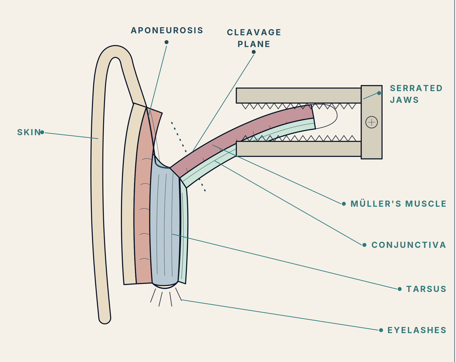
The eyelid is everted over a Desmarres retractor. A marking suture lifts the conjunctiva and Müller’s muscle above the upper tarsal border, the clamp closes on that raised fold, and the retractor is withdrawn by rotation. An absorbable suture is run as a U-shaped mattress just distal to the jaws, and the tissue held above it is resected. The tarsus never entered the jaws at all.
The arithmetic that catches people out
This is the detail the instrument imposes and that quick descriptions skate over. The clamp does not take a height of tissue, it takes a fold. The raised tissue passes twice between the jaws.
Put another way: for a total resection of 6 to 10 millimetres, the marking goes 3 to 5 millimetres above the upper tarsal border, not at the height you mean to resect. A surgeon who marks at the target height resects twice what they intended — and on an eyelid, doubling the error is not something you retrieve with a suture.
Patient selection is less clear-cut than it reads
Custom has it that phenylephrine is instilled into the superior fornix and that the patient is held back if the lid lifts. A brisk response is reassuring, and the test remains a reasonable instrument of preoperative discussion.
But it does not deserve the status of verdict that is sometimes lent to it. Its predictive value has been questioned, and published series report good results in phenylephrine-negative patients simply by increasing the resection — five millimetres where four would have been done. The test informs the decision; it does not take it in place of the surgeon, who remains the sole judge of levator function, degree of ptosis and tarsal integrity.
Today
The clamp is becoming optional. Single-suture variants, suture-less mullerectomies and needle techniques reproduce the same resection without a dedicated instrument. It is possible that the Putterman clamp will end up where it had meant to send the Fasanella-Servat — except that the Fasanella-Servat outlived its own obituary.
What will remain is not the instrument. It is the order of operations: assess the eyelid, decide what it needs, and only then choose the tool. Never the other way round.
Techniques pass. The discipline that grounds the indication endures.
References
- Fasanella RM, Servat J. Levator resection for minimal ptosis: another simplified operation. Arch Ophthalmol. 1961;65:493-496.
- Putterman AM. A clamp for strengthening Müller’s muscle in the treatment of ptosis. Modification, theory, and clamp for the Fasanella-Servat ptosis operation. Arch Ophthalmol. 1972;87(6):665-667.
- Putterman AM, Urist MJ. Müller muscle-conjunctiva resection. Technique for treatment of blepharoptosis. Arch Ophthalmol. 1975;93(8):619-623.
- Dresner SC. Further modifications of the Müller’s muscle-conjunctival resection procedure for blepharoptosis. Ophthalmic Plast Reconstr Surg. 1991;7(2):114-122.
- Baldwin HC, Bhagey J, Khooshabeh R. Open sky Müller’s muscle-conjunctival resection in phenylephrine test-negative blepharoptosis patients. Ophthalmic Plast Reconstr Surg. 2005;21(4):276-280.
- Leung VC, Dupuis JEK, Ashraf DC, et al. Müller muscle conjunctival resection: a multicentered prospective analysis of surgical success. Ophthalmic Plast Reconstr Surg. 2023;39(3):226-231.